 Muscle Fibres Part 3
 Index
to Course Material
Index
to Course Material Index
to Section 12
Index
to Section 12 Part
2
Part
2 References
Myofibril Contraction
Upon contraction of a myofibril, the "walking" of the myosin heads along
the thin filaments means that the overlap of the two types of filament,
i.e. the width of the AI band, increases; this is at the expense of the
AH zone, so that the total width of AI and AH (i.e. the length of a myosin
thick filament) is invariant. As the length of the thin filaments is also
constant, the increase in the width of AI is matched by a decrease in that
of I.
The Contraction Cycle
In relaxed muscle, the S1 heads of the myosin molecules of the thick filaments
are detached from the thin filaments, and orientated perpendicular to them.
One molecule of ADP and one Pi (phosphate) group are bound to the
myosin head.
The contraction cycle can be described in 4 stages:
-
Stimulation of the muscle results in the S1-ADP-Pi complexes binding
to the adjacent thin filaments, still perpendicular to them.
-
The interaction between myosin and actin results in the release of Pi,
followed by ADP, which induces a conformational change in the myosin molecules:
hinge bending tilts the head through approximately 45°. This motion
pulls the thin filaments approximately 100Å towards the M line, in
a "rowing" action: thus the "power stroke".
-
Binding of ATP to the S1 heads causes them to detach from the thin filaments,
still in the tilted conformation.
-
The bound ATP is hydrolyzed (see below), returning the S1 head to the former
relaxed conformation.
 Diagram. (11Kb)
During the contraction of a muscle, this cycle occurs many times as
the myosin heads walk along the thin filaments; the length of a contracted
muscle may be as little as two-thirds of its fully extended state.
The ATP hydrolysis in Step 4 above is carried out by myosin itself
(the globular S1 heads are ATPases; see Part 1.)
In solution studies, the turnover number of this reaction is found to be
increased (by a factor of 200) by actin, by means of accelerating the release
of ADP and Pi from the actomyosin complex (step 2); the hydrolysis
step itself is carried out rapidly by myosin alone. Magnesium ions are
required for these reactions. Addition of ATP to a solution of the complex
is found to decrease the affinity of actin for myosin; this corresponds
to step 3.
 A movie of the contraction cycle
is available in the following formats:
A movie of the contraction cycle
is available in the following formats:
N.B. The power stroke is not shown to scale. In these movies,
two power strokes (approx 100Å) move the thin filament by one turn,
whereas each turn of such a filament is 360Å in length
The role of Troponin and Tropomyosin
Thin filaments consist of actin filaments with one troponin-tropomyosin
complex for each 7 actin monomers; refer to the section in the previous
chapter .
-
Actomyosin complexes obtained from purified actin and myosin exhibit
contraction upon the addition of ATP.
-
Actomyosin complexes prepared from muscle tissue (which therefore
include tropomyosin and troponin in the thin filaments) do not contract
upon the addition of ATP, unless calcium ions are present.
This indicates that the troponin-tropomyosin complex regulates muscle contraction
in response to the levels of Ca²+ ions. Only the troponin subunit
TnC binds Ca²+.
An allosteric mechanism is believed to regulate the binding of myosin
to actin, and thus muscle contraction. In the relaxed state, the tropomyosin
molecule binds along the groove in the actin double helix, and blocks the
S1-binding sites of the seven actin monomers. Binding of Ca²+ to troponin
C causes a conformational change; interaction between troponin and tropomyosin
moves the latter approximately 10Å deeper into the groove, exposing
the myosin-binding sites. Refer to Zot and Potter (1987).
The Ca²+ ions are delivered from the lumen of the sarcoplasmic
reticulum, a network of flattened membrane-bound sacs which surrounds
all the myofibrils in a muscle cell; the membrane is made temporarily permeable
to Ca²+ ions upon the arrival of a nerve impulse.
Index
to Course MaterialIndex
to Section 12Part
2References
Last updated 16th Jul '96
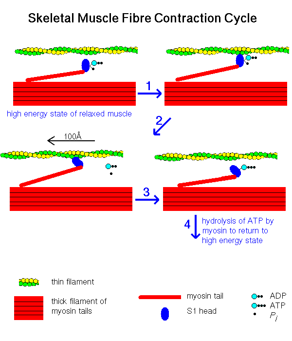 Diagram. (11Kb)