 Muscle Fibres Part 1
 Index
to Course Material
Index
to Course Material Index
to Section 12
Index
to Section 12 Part
2
Part
2
Introduction
This section describes an example of how the form of structural proteins
relates to motion: namely the contraction of skeletal muscle .
Other examples to investigate include:
-
the beating of cilia and flagella, which are based on
microtubules
-
the movement of chromosomes, the precise coordination of which is
essential during cell division; this also involves microtubules
-
cell movement and determination of cell shape- many aspects of which
involve interactions of membrane proteins with the extracellular
matrix (composed of glycoproteins,
fibrous proteins, polysaccharides)
(Here are links to the course pages dealing with
microtubules
,
membrane proteins ,
fibrous
proteins )
Skeletal muscles (also called striated or striped muscle)
are only one type of muscle tissue occurring in vertebrates. They are generally
under voluntary control. The other two are (i) cardiac (i.e. heart)
muscle, which is specialized but resembles skeletal muscle in many respects,
and (ii) smooth muscle which is generally controlled involuntarily
by the autonomic nervous system .
The Structure of Skeletal Muscle
Muscles, Myofibres and Myofibrils
Skeletal muscle cells are highly specialized. They are called myofibres,
cylindrical in shape, 0.01 - 0.05 mm in diameter and 1 - 40 mm long. These
multinucleate
cells consist of a bundle of myofibrils surrounded by a plasma membrane.
A muscle consists of a bundle of myofibres.
Myofibrils
A myofibril consists of repeating identical units called sarcomeres.
Regular repeating formations of two types of protein filaments are the
basis of the sarcomere:
-
thin filaments- these consist mostly of
actin , with tropomyosin
and troponin; these have already been described in an
earlier chapter.
-
thick filaments -composed of the protein myosin
The structure of the sarcomere is described on the next page.
Structure of Myosin
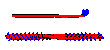
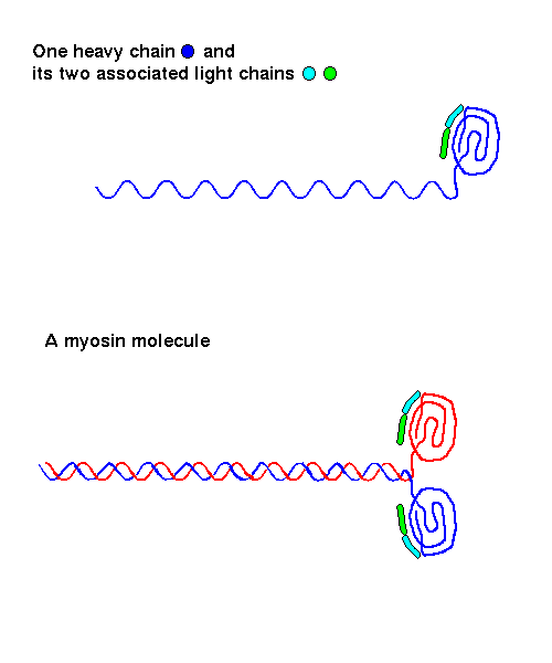
A single myosin molecule consists of two heavy chains and four light chains.
It is effectively a dimer of two heterotrimers, each of which consists
of two different light chains (approximately 20 kD in mass) and a single
heavy chain (230 kD). The latter has a globular head and a long alpha-helical
tail. In fact the two tails of the complete molecule form a parallel coiled-coil,
so that myosin consists of a long (1500 Å) fibre, 20 Å thick,
with a two-headed globular end. There is a hinge region between each head
and the tail section.
 Here
is a diagram.
(5Kb)
Here
is a diagram.
(5Kb)
A myosin molecule has three functions:
-
binding to other myosin molecules to form filaments; this occurs spontaneously
in physiological conditions. At high ionic strengths, myosin exists as
individual molecules.
-
binding to actin filaments
-
it is an ATPase, i.e. it hydrolyzes ATP to give ADP and a phosphate
(Pi).
The two different structural domains are responsible for different functions,
as is revealed by treatment of myosin with proteases to give different
subunits. Cleavage of myosin with trypsin gives two products:
-
light meromyosin (LMM), an 850Å coiled-coil, i.e. a large
section of the myosin "tail". LMM aggregates to form filaments, but does
not bind to actin filaments, and does not hydrolyze ATP.
-
heavy meromyosin (HMM), which consists of the globular heads and
a shorter section of tail. It does not aggregate to form filaments, but
it hydrolyzes ATP and binds to thin filaments.
Treatment of HMM with the protease papain cleaves the two globular heads
from the tail section (S2). The two heads (termed S1) are
not surprisingly found to be the site of ATPase activity; they also bind
to actin filaments.
 Click here for the crystal structure (C-alpha
atoms only) of a proteolytic fragment of myosin from chicken muscle. 1mys
(100Kb) [Bbk|BNL|ExP|Waw|Hal]
Click here for the crystal structure (C-alpha
atoms only) of a proteolytic fragment of myosin from chicken muscle. 1mys
(100Kb) [Bbk|BNL|ExP|Waw|Hal]
This fragment consists of an entire S1 head (843 residues) and two light
chains.
Diagram. (16Kb) To achieve this rendition, colour
the molecule by 'chain' and select 'spacefill' from the menu.
This fragment (all atoms) contains two light
chains and 60 residues of the heavy chain.
1scm (238Kb) [Bbk|BNL|ExP|Waw|Hal]
Diagram. (12Kb)
Myosin is therefore unusual in that it is both a fibrous protein, and
a globular enzyme.
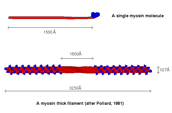
Myosin filaments
Thick filaments consist almost entirely of myosin. A myosin filament contains
several hundred myosin molecules in two bundles packed end to end. In each
bundle, the myosin molecules all point in the same direction, giving an
aggregation of the myosin tails with the globular heads protruding in a
regular helical arrangement. The two bundles are packed such that the ends
of the tails are facing each other, but the tails from one bundle overlap
those of the other, so that the "bare zone" where there are no S1 heads
is less than twice the length of a single myosin tail.
 Here is a diagram.
Index
to Course MaterialIndex
to Section 12Part
2
Last updated 16th Jul '96
 Here
is a diagram.
(5Kb)
Here
is a diagram.
(5Kb)
 Here is a diagram.